Brain Experience.
A journey towards the inner self of your brain, brought to you by Baihan Lin.
Please press the right-left key to navigate the journey.
Introduction
Through the eye of a cognitive neuroscientist
Cognitive neuroscience is the research field that studies the biological processes and psychological aspects that underlie cognition, with a specific focus on the neural connections in the brain which are involved in mental processes. There are many things cognitive neuroscientist do: we might study how the brain understands language, music, do math, or think creatively. In this journey, we are going to focus on brain mechanisms behind the perception, and more specifically, visual perception. Wanna join us? Buckle up.
Background
This is the Way...
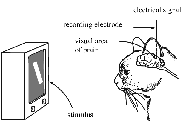
Usually, neuroscientists first define a research question (e.g. how do the background noise affect how different brain regions perform their functional role in categorizing one object from another?) Then, we design controlled experiments with specific conditions that test the hypothesis (e.g. images with different background noise as the visual stimuli). We recruit participants to perform these tasks while we record their brain activities. Finally, we analyze the data and arrive at a explanation.
Experiments
• Psychophysics
• Event-related potential
• Lab on a chip
Data collection
• Neuroimaging
• Neuronal recording
• Single-cell techniques
Analysis
• Functional connectivity
• Brain-wide association analysis
• Time series analysis
Inference
• Representational similarity analysis
• Deep neural networks
• Theoretical modeling
Method
Into our visual perception problem.

We want to study major visual cortical areas in the brain and how they operate when
viewing scenes of naturalistic conditions.
To investigate this question, we collect brain activity data from the subjects when they
are viewing a series of images. In this case, to examine the cellular responses to natural
stimuli, the images being used consist of a library of 118 natural scenes, selected from
three different databases (Berkeley Segmentation Dataset, van Hateren Natural Image Dataset
and McGill Calibrated Colour Image Database). A scene image was presented briefly (250 ms)
then replaced with the next scene image. Each image was presented 50 times, in a random order,
with intermittent blank intervals.
We study multiple brain regions at the same time.
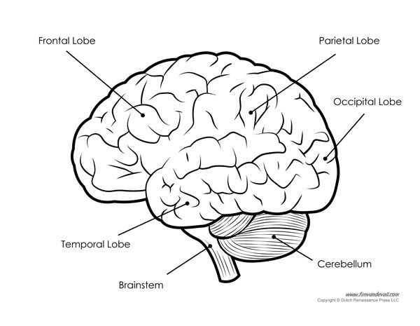
To understand the function of different brain regions, we use a neuroimaging technique, called calcium imaging, to measure the activity pattern of different brain regions of interest (ROIs). To be more precise, we are interested in the perceptual behavior of six visual brain areas: primary visual cortex (V1), laterointermediate (LM), posteromedial (PM), rostrolateral (RL), anteromedial (AM), and anterolateral (AL) visual areas.

Visualization
What are the teams at play when the brain sees an image?
This is what the data looks like. Please click on the visualization to activate the
interactive sequence.
What we are seeing now, is the signal value of many neurons recorded from the study where multiple participants view different images at different time points, colored by their brain regions.
Next steps
How does the brain work?
A fundamental challenge for cognitive and systems neuroscience is to quantitatively relate its three major branches of research: brain-activity measurement, behavioral measurement and computational modeling. And to learn how cognition is implemented in the brain, we must build computational models that can perform cognitive tasks, and test such models with brain and behavioral experiments.

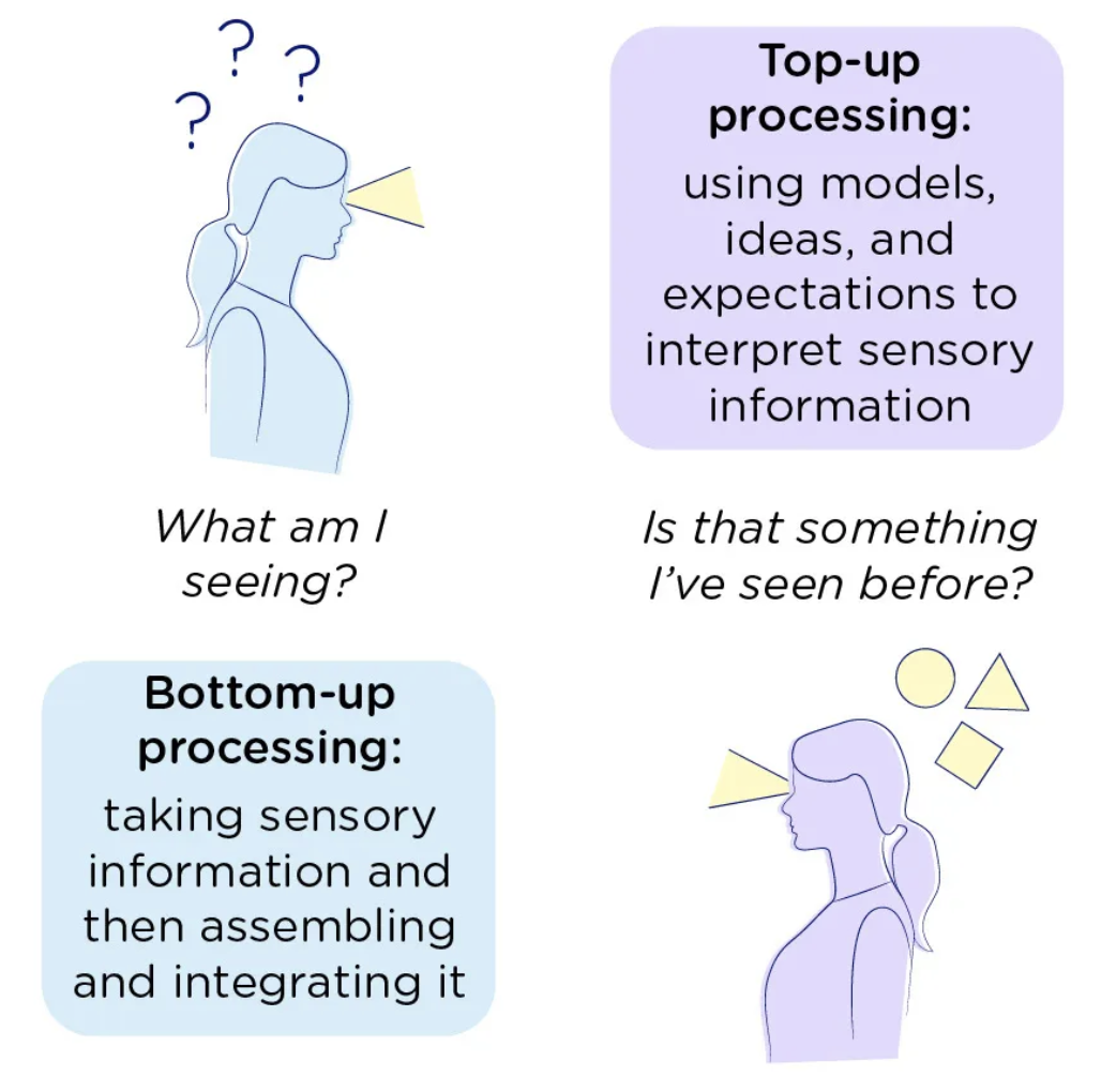
Even in the subproblem of the visual processing, here are various mechanisms and dimensions that are the topics of active research, waiting to be gradually uncovered.
From the perception to cognition, there are also more layers of subtlety, such as the attention mechanisms and contextual information in real world scenarios.
